
Carolyn Lou
Automated Detection of Paramagnetic Rims in Multiple Sclerosis Lesions on 3T Susceptibility-based MR Imaging
Presenter
Flash Talk PresenterI am a doctoral candidate in biostatistics at the University of Pennsylvania as a part of the Penn Statistical Imaging and Visualization Endeavor (PennSIVE) Center. My research interests include statistical and computational methods for the analysis of neuroimaging data and clinical trials.
Abstract
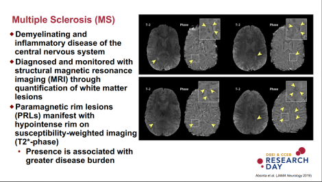
The presence of a paramagnetic rim around a white matter lesion has recently been shown to be a hallmark of a particular pathological type of multiple sclerosis (MS) lesion. Increased prevalence of these paramagnetic rim lesions (PRLs) is associated with a more severe disease course, but manual identification of PRLs is time-consuming. We present a method to automatically detect PRLs on 3T T2*-phase images. T1-weighted, T2-FLAIR, and T2*-phase MRI of the brain were collected at 3T for 19 subjects with MS. Images were then processed with automated lesion segmentation, lesion labelling, and lesion-level radiomic feature extraction. A total of 877 lesions were identified, 118 (13%) of which contained a paramagnetic rim. We fit a random forest classification model on a training set and assessed our ability to classify PRL lesions on a test set. The number of PRLs per subject identified via our automated lesion labelling method was highly correlated with the gold standard count of PRLs per subject, r = 0.91 (95% CI [0.79, 0.97]). The classification algorithm achieved an area under the curve of 0.80 (95% CI [0.67, 0.86]).
Keywords
neuroimaging; radiomic image analysis; machine learning;Commenting is now closed.
About Us
To understand health and disease today, we need new thinking and novel science —the kind we create when multiple disciplines work together from the ground up. That is why this department has put forward a bold vision in population-health science: a single academic home for biostatistics, epidemiology and informatics.
© 2023 Trustees of the University of Pennsylvania. All rights reserved.. | Disclaimer